NIH: The retina, like this one from a mouse that is flattened out and
captured in a beautiful image, is a thin tissue that lines the back of
the eye. Although only about the size of a postage stamp, the retina
contains more than 100 distinct cell types that are organized into
multiple information-processing layers. These layers work together to
absorb light and translate it into electrical signals that stream via
the optic nerve to the brain. In people with inherited disorders in which the retina degenerates,
an altered gene somewhere within this nexus of cells progressively robs
them of their sight.
Only good, independent and reliable information about health from experts.
Showing posts with label retina. Show all posts
Saturday, November 12, 2016
Zika Virus Can Cause Severe Damage to Retina in Infants
JAMA: In a study published online by JAMA Ophthalmology, Rubens
Belfort Jr., M.D., Ph.D., of the Federal University of Sao Paulo,
Brazil, and colleagues examined the affected retinal layers in infants
with congenital Zika syndrome and associated retinal abnormalities using
optical coherence tomography (OCT). The study included 8 infants (age range, 3-5.1 months) with
congenital Zika syndrome (CZS), the term created for a variety of
anomalies associated with intrauterine Zika virus infection. Optical
coherence tomographic images (a noninvasive diagnostic imaging tool that
provides cross-sectional retinal images) were obtained in the affected
eyes of 7 infants with CZS who had undergone previous ophthalmologic
examinations on March 17, 2016, and in 1 infant on January 1, 2016. An
IgM antibody-capture enzyme-linked immunosorbent assay for Zika virus
was performed on the cerebrospinal fluid samples of 7 of the 8 infants,
and other congenital infections were ruled out.
Sunday, November 6, 2016
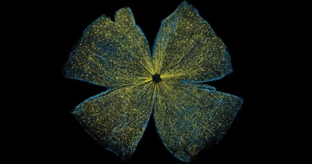
Artificial retinas : promising leads towards clearer vision
CNRS: A major therapeutic challenge, the retinal prostheses that have been under development during the past ten years can enable some blind subjects to perceive light signals, but the image thus restored is still far from being clear. By comparing in rodents the activity of the visual cortex generated artificially by implants against that produced by “natural sight”, scientists from CNRS, CEA, Inserm, AP-HM and Aix-Marseille Université identified two factors that limit the resolution of prostheses. Based on these findings, they were able to improve the precision of prosthetic activation. These multidisciplinary efforts, published on 23 August 2016 in eLife, thus open the way towards further advances in retinal prostheses that will enhance the quality of life of implanted patients.
Monday, February 25, 2013
Age-related macular degeneration (AMD) Overview
Source: Plos Medicine. 2013.
The third leading global cause of blindness (after cataracts and glaucoma) is age-related macular degeneration (AMD).
This group of conditions is characterized by lesions in the macular (central) region of the retina, the tissue at the back of the eye that converts light into electrical messages and sends them to the brain.
AMD, which affects older people, destroys the sharp central vision that is needed for reading or driving, leaving only dim, blurred images or a black hole at the center of vision.
AMD can be diagnosed by examining digital photographs of the retina or by examining the retina directly using a special magnifying lens (slit lamp biomicroscopy).
There is no cure for AMD, although injections into the eye of certain drugs, such as bevacizumab, that block the activity of vascular endothelial growth factor can slow the rate of vision loss caused by some forms of AMD.
Subscribe to:
Comments (Atom)

