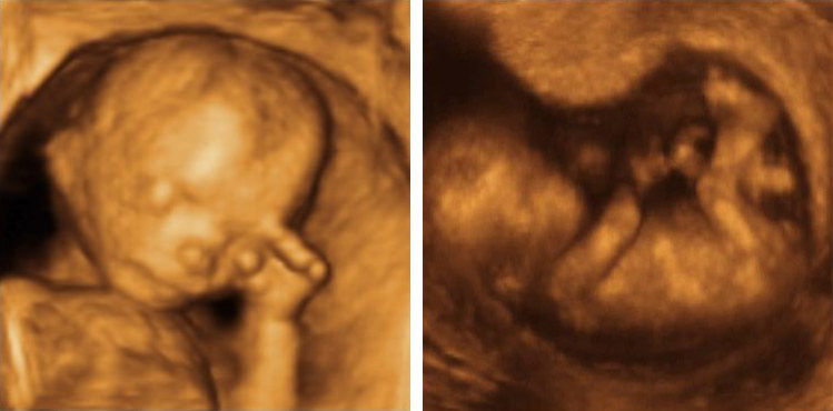
The object under observation is scarcely larger than a thumb nail. Three times a second it contracts and presses a few drops of blood through the 18-week-old embryo. Fifteen centimetres of skin, fat and muscle tissue separate it from the eye of the observer. And yet: on the computer screen a three-dimensional image of the baby's heart pulsates which is astonishingly accurate in its details. When Dr. Ulrike Herberg, child cardiologist at the University of Bonn, uses the mouse to turn it in the appropriate direction, even a layperson can recognise the cardiac valves opening and closing.
Ultrasonic diagnostics is usually brainwork: "Conventional equipment gives us two-dimensional images which are cross-sections, e.g. cutting through the ventricles," Dr. Herberg explains. What the ventricles look like spatially, whether they are smaller than normal or perhaps are defective in the way they contract, has to be resolved by the doctors by assembling these cross-sections in their minds into a 3D model. "For this even with an organ which does not move an excellent sense of space and plenty of experience are required, and this is even more necessary with the heart, which regularly contracts and then fills up with blood again." The best ultra-sound specialists can thus say with more than 80% certainty whether the unborn child is suffering from a heart defect - less experienced medical personnel often only score a success rate of 25%.
Dr. Ulrike Herberg is at present developing a method which is meant to facilitate this mental exploit in conjunction with the University of Bonn's Clinic for Obstetrics and Pre-natal Diagnostics and experts of the software company MedCom Ltd. It involves the doctors placing a conventional ultra-sound probe onto the pregnant patient's stomach and swivelling it in such a way that takes in the complete heart of the unborn baby within 20 seconds. During this time the machine records about 1000 images. At the same time it records the foetus's heartbeat with a special sensor. This is the "baton" which tells the computer which images belong to which "part of the beat". For example, if the baby's heart has contracted and expanded again 60 times during the examination, the machine has recorded a total of 60 cross-sections during its swivelling movement at the time of maximum contraction, which are all taken from different areas of the heart. The imaging software can now assemble a composite 3D picture from these two-dimensional ultra-sound images. When the probe is being swivelled for the maximum of 20 seconds the software receives 3D pictures by this means from different phases of the heartbeat - from complete contraction to complete relaxation. Thus the entire movement of the myocardium can be followed on the screen. "We can even see exactly how the cardiac valves open," Dr. Herberg enthuses, adding: "information which is not available from the conventional cross-sections." An additional advantage is that the three-dimensional pictures can be turned whichever way is needed, so that the surgeon can see the operating field on the screen and plan the therapeutic strategy better.
Some cardiac diseases might perhaps be treated or prevented before birth if detected early enough - "a perspective which I find particularly attractive," Dr. Herberg declares. Thus the software enables the volume of the heart to be calculated much more precisely than in the past - often an early indicator that something is going wrong in the development of the cardiac muscle. Twins, for instance, may develop a joint cardiovascular system via the placenta. "The one twin then keeps pumping blood into its sister's or brother's bloodstream, thereby leading to the second twin's heart being overloaded: it becomes excessively large, with serious heart defects being the possible consequences." With early detection, however, this type of malformation can be prevented by firing a laser at the joint blood vessels and thereby sealing them.
Minor defects still need to be tackled by the designers: the measurement of the heartbeat by the special sensor, for examaple, can reduce the quality of the images, and the art of swivelling the probe evenly needs practice. Once these teething troubles are over, the method must show in clinical trials if it really is an improvement on conventional ultra-sound diagnostics.
Contact person for the media: Dr. Ulrike Herberg, Paediatric Centre of the University of Bonn, Tel.: ++49-228-2873256, e-mail: ulrikeherberg@hotmail.com