This has led to a number of human clinical trials—with some encouraging progress being reported for at least one condition, Leber congenital amaurosis—that are transferring a normal version of the affected gene into retinal cells in hopes of restoring lost vision.
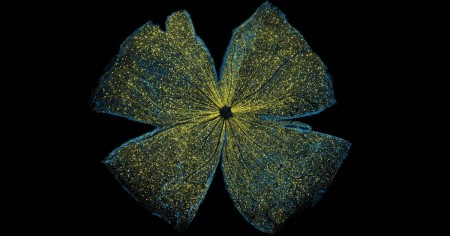
To better understand and improve this potential therapeutic strategy, researchers are gauging the efficiency of gene transfer into the retina via an imaging technique called large-scale mosaic confocal microscopy, which computationally assembles many small, high-resolution images in a way similar to Google Earth. In the example you see above, NIH-supported researchers Wonkyu Ju, Mark Ellisman, and their colleagues at the University of California, San Diego, engineered adeno-associated virus serotype 2 (AAV2) to deliver a dummy gene tagged with a fluorescent marker (yellow) into the ganglion cells (blue) of a mouse retina. Two months after AAV-mediated gene delivery, yellow had overlaid most of the blue, indicating the dummy gene had been selectively transferred into retinal ganglion cells at a high rate of efficiency [1].
The researchers also used AAV2 to deliver into the retinas of mice a gene that coded for a mutant version of a protein, called DRP1. They found it inhibited normal DRP1 protein and stopped retinal ganglion cells from dying in response to glaucoma, a vision-threatening condition that elevates pressure within the eye. The gene transfer proved successful in rescuing the retinal ganglion cells, and researchers are continuing to pursue this line of study with the aim of translating their discoveries into possible ways of helping humans with glaucoma.
It’s also worth noting that this image recently took top honors in the NIH Institute and Centers Art Challenge, which was held in October as part of the Combined Federal Campaign, in which federal employees make their annual contributions to charitable organizations. The art competition had a number of outstanding entrants, some of which will be on display during November at the NIH Clinical Center in Bethesda, MD. If you’re in the neighborhood, come take a look!
References:
[1] DRP1 inhibition rescues retinal ganglion cells and their axons by preserving mitochondrial integrity in a mouse model of glaucoma. Kim KY, Perkins GA, Shim MS, Bushong E, Alcasid N, Ju S, Ellisman MH, Weinreb RN, Ju WK. Cell Death Dis. 2015 Aug 6;6:e1839.