Cholangiocarcinoma (CCA) is a biliary tract cancer (a cancer that starts in a bile duct) originating in the epithelium of the biliary tree, either intra or extra hepatic.
Who gets cholangiocarcinoma?
The frequency is unknown but it accounts for approximately 3% of gastrointestinal tumors and 10-15% of all hepatobiliary cancers (liver, or bile duct or gallbladder).
Cholangiocarcinoma can occur in the intra or extra-hepatic biliary tract (inside or outside the liver).
A specific type of extra-hepatic CCA known as a Klatskin tumor occurs at the junction where the left and right hepatic bile ducts meet the common bile duct (CBD). It is slightly more prevalent in males than females (1.3:1.0) and usually presents in the fifth to seventh decade of life.
Symptoms
Clinical manifestations are not usually noted until an advanced disease stage. Extra-hepatic CCA manifests with signs of cholestasis (jaundice, pale stools, dark urine, pruritus / itching), malaise, weight loss and/or progressive weakness. Intra-hepatic CCA may present with an abdominal mass or with non-specific symptoms of decreased appetite, weight loss, abdominal pain and malaise.
Causes
The causes are unknown and most cases of CCA occur sporadically.
Risk factors include primary sclerosing cholangitis an inflammatory condition of the biliary tree, secondary sclerosing cholangitis, chronic typhoid carriage, parasitic infections (Opisthorchis viverrini and Clonorchis sinensis), exposure to thorotrast (x-ray contrast medium) and choledochal cysts, all of which cause chronic biliary inflammation.
Gallstones and cholecystectomy are not thought to be associated with an increased frequency of cholangiocarcinoma.
Diagnosis
- Diagnosis is suspected on clinical and laboratory findings.
- Serum carbohydrate antigen (CA 19-9) is the glycoprotein tumor marker most often used in the diagnosis of CCA. It is found to be elevated in 85% of patients.
- Increased CEA levels (another tumor marker) are also noted.
- Extra-hepatic tumors cause increased levels in the blood of alkaline phosphate, conjugated bilirubin and gamma-glutamyl transpeptidase while intra-hepatic have only slightly elevated alkaline phosphate levels.
- Abdominal imaging, visualization of the biliary tree and biopsies of the lesion are necessary for diagnosis.
- Magnetic resonance cholangiopancreatography (MRCP) provides information on intrahepatic metastases, biliary anatomy and tumor extension and is used in the staging of CCA. It has been advocated to replace endoscopic retrograde cholangiopancreatography (ERCP), an endoscopic more invasive method.
- Visualization of the biliary tree and samples through brush cytology or bile duct biopsies are obtained with ERCP (endoscopy). see picture below
- A needle biopsy is performed in those with a liver mass.
- Extra-hepatic CCA is further divided into anatomical subtypes according to the Bismuth classification and a disease stage is given.
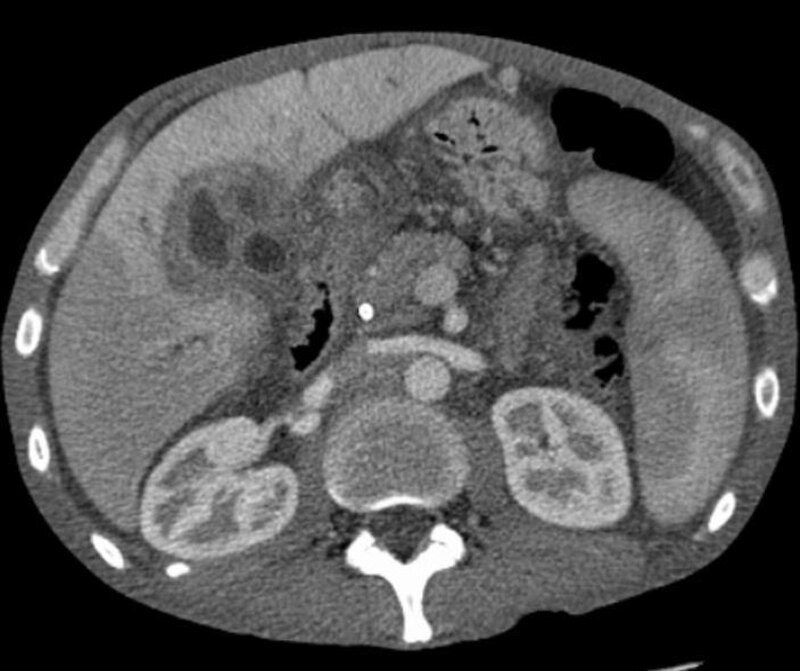
- Ultrasound, and contrast enhanced helical computerized computed tomography (CT) can be used in visualizing the extent of disease.
- Intra-hepatic CCA is often mistaken for metastatic adenocarcinoma. Carcinoma of the gallbladder, benign strictures and Mirizzi syndrome should be excluded.
 |
| ERCP of cholangiocarcinoma |
Treatments
Surgical resection is the only potentially curative treatment for CCA but recurrences after surgery are frequent. Unfortunately CCA is often diagnosed as unresectable because of local extension and/or metastases. Distal CCA arising from the CBD is often treated by pancreatoduodenectomy. More proximal CCA needs hepatic resection.
Palliative management involves biliary drainage by inserting metal stents in the biliary tree to release the blockage.
Adjuvant chemotherapy after surgery or palliative chemotherapy for unresectable CCA is indicated. Gemcitabine combined with cisplatin therapy is the standard treatment for unresectable biliary tract cancers, including CCA.
As proximal CCA is usually not diagnosed until a late stage of disease, prognosis is poor with 5-year survival rates of 20-50% after resection and almost 0% in unresectable tumors. Death is often due to biliary sepsis, cancer cachexia, malnutrition and liver failure.