Author :
David J. Hackam, MD, PhD
Attending Pediatric Surgeon, Children’s Hospital of Pittsburgh
Associate Professor of Surgery
University of Pittsburgh School of Medicine
Co-Director, Fetal Diagnosis and Treatment Center, Children’s Hospital of Pittsburgh
2008-07-28
2008-07-28
Highlights
- Necrotizing enterocolitis (NEC) is the leading cause of death from gastrointestinal disease in neonates (newborn babies)
- The pathological manifestations of NEC include patchy inflammation leading to full thickness necrosis and perforation of the intestine
- Treatment of NEC involves cardiorespiratory support, broad spectrum antibiotics, and surgical resection of necrotic intestine
- In mild to moderate cases of NEC, the outcome is very good; In severe cases, mortality rates are high, and long-term morbidity is common
1. The difficult problem that is necrotizing enterocolitis.
Ask
any practicing neonatologist or pediatric surgeon to describe the most
vexing and frustrating problem that they face as they take care of
newborn patients, and they are likely to give you the same answer –
“necrotizing enterocolitis.” This is a disease that in many ways is a
product of our success as caregivers to those born too soon, a disease
that attacks predominantly (although by no means exclusively) preterm
infants, a disease that strikes seemingly without warning, always
without regard for the fragility of the tiny host. Necrotizing
enterocolitis – or NEC as it is usually abbreviated – is a disease that
attacks the intestines from within, and transforms them into a necrotic
(dead tissue), septic stew that if untreated progresses to multi-system
organ failure and death in over a third of patients. In certain cases,
surgery may be indicated, in which the abdomen may be drained and/or the
necrotic bowel may be removed, potentially leaving the tiny patient
with inadequate intestine to support his or her nutritional needs. As
for the families of children with NEC, they are hurt in two ways: first,
through the pain and suffering of having a tiny child face such a
devastating disease, and second, through the almost universal lack of
even the remotest awareness as to what NEC actually is.
2. What is necrotizing enterocolitis?
Necrotizing
enterocolitis (NEC) is the leading cause of death from gastrointestinal
disease in preterm neonates, and will likely soon overtake respiratory
disease as the leading case of death overall in these patients. NEC is
diagnosed in between 0.9 to 2.4 per 1000 live births, and the increase
in survival rates of premature infants have led to an overall increase
in the incidence of this disease 1-4. NEC is both an acute
and chronic disorder that is characterized initially by intestinal
inflammation, yet may progress to intestinal necrosis, with perforation
in more advanced cases. In the most severe form, NEC may lead to
overwhelming multi-system organ failure and death from systemic sepsis.
NEC may indeed be considered as a spectrum of diseases. At one end of
the spectrum, patients with NEC may have relatively mild symptoms that
are readily responsive to medical treatment. At the other end of the
spectrum, patients may develop a fulminant course, which is
characterized by near total destruction of the entire intestine. The
recognition of the fact that patients with NEC exist along a wide
spectrum of presentations facilitates a greater understanding of the
mechanisms that lead to its development, as well as strategies that may
be most appropriate to treat it.
3. Baby Jimmie – an infant with NEC.
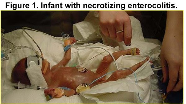
NEC
is a disease that initially affects the gastrointestinal system, then
progresses to lead to the development of multi-system organ failure in
certain cases. A baby with NEC is shown in Figure 1 – note his
small size in comparison with the nurse’s hands, the numerous monitors
and tubes to which he is attached, and the swollen distended abdomen. In
order to more clearly illustrate the clinical presentation of NEC,
consider the following description of baby Jimmie (NB: for those of you
that don’t like unhappy endings, you may want to skip ahead. But that’s
NEC.)
After
five years of trying, and having almost given up entirely on their
chances, Rhonda and James were finally pregnant. Both in their early
forties, they were wondering if being first time parents was such a good
idea after all anyway. But they so desperately wanted a child that they
didn’t dwell on anything but their immense desire to raise a family.
The first trimester couldn’t go away fast enough, but Rhonda settled
into the idea of being pregnant so that by the time the second trimester
arrived, she started to plan for the future. And when she first felt
tiny Jimmie (they had decided on that name before even knowing that they
were having a boy) move, it was as if her whole life had been
transformed. On her ultrasound at 24 weeks they actually came face to
face (or face to ultrasound probe) with their tiny child, and started to
bond with him in a way that only expectant parents can ever understand.
And then, a month after her ultrasound, when Rhonda was just seven
months pregnant, a bad thing happened. Rhonda went into labor. At first
she didn’t believe it – she thought the fluid trickling down her inner
leg must just be normal discharge; she hoped that the pain in her lower
abdomen was indigestion. But when the pain worsened and
the fluid gushed, she knew that she was going to meet Jimmie face to
face much sooner than anyone had predicted.
Rhonda
and James rushed to the hospital, where a fetal assessment indicated
that their baby was sick, that his heart rate had become irregular, that
he needed to be delivered. And so, barely an hour after the onset of
labor, little Jimmie was born – a tiny child, precious and fragile and
beautiful. Unable to breathe on his own, Jimmie was intubated and placed
on a ventilator. For the first week of his life, Jimmie seemed to do
pretty well, and the doctors stated that he would just need to feed and
grow and would likely be fine. And so they started to feed him –
dripping infant formula into a little tube that was passed into Jimmie’s
nose. And while Jimmie seemed to like the formula, his body clearly did
not. Two days after the feedings had started, Rhonda and James came to
Jimmie’s bedside to see that his abdomen had become swollen, his
previously warm skin was now cold and clammy, and he looked like he was
dying. An x-ray revealed that his intestines were sick – a disease the
doctors called necrotizing enterocolitis – a disease that Rhonda and
James had never heard of. Jimmie needed urgent surgery,
where a portion of his intestines were removed, leaving him with two
tiny pieces of separate intestine extruding through his skin – temporary
stomas, as the doctors called them. Little Jimmie began to improve, and
a few weeks later underwent another operation to reattach his
intestine. He was now starting to tolerate feeds, and was starting to
grow, to interact, to babble to laugh. But the effects of NEC never went
away. Jimmie had insufficient bowel length to absorb his formula – a
condition that the doctors called “short bowel syndrome.” Dependent upon
being fed through a vein, little Jimmie began to get worse. He suffered
infection after infection, then a slow, steady, downward spiral. At the
age of six months of age, his little body unable to keep up with ever
increasing infections, he died with his parents at his side.
4. Risk factors for the development of NEC.
Based
on several large clinical series, several risk factors have been
implicated in the development of NEC. The most common factors include
prematurity and aggressive administration of enteral feeds. Other risk
factors for the development of NEC include episodes
of birth asphyxia, umbilical vessel catheterization (as is often
required for monitoring and infusion of fluids in small infants),
African American descent, and congenital heart disease. In addition,
there are maternal factors that lead to the development of NEC,
including maternal cocaine use and maternal pre-eclampsia.
By contrast, the major protective factor in NEC that has been
consistently revealed in a large series of studies is the administration
of breast milk 5. These factors indicate that NEC typically develops in the setting of a stressed, formula-fed, preterm infant.
5. How is NEC diagnosed?

NEC
is typically diagnosed on the basis of a combination of clinical,
radiographic, and laboratory features, and is observed along a spectrum
of disease severity. In order to understand the modes of presentation of
NEC, and for the purposes of the current discussion , several scoring
systems for the diagnosis have been devised that grade disease severity
based upon the presenting symptoms and evaluations. The most commonly
used classification system for NEC was described by Bell and colleagues
in 1978 and remains commonly used today. Specifically, Bell and
colleagues characterized the severity of NEC as Stage I (mild), Stage II
(moderate) and Stage III (severe, see Figure 2 and reference 6). In all cases, the diagnosis of NEC and the particular grade of NEC is established using a combination of clinical and radiographic findings.

In
mild cases (Bell Stage I), infants demonstrate difficulty breathing
(apnea, nasal flaring, retractions). There may be episodes of heart rate
irregularities (bradycardia, tachycardia), as well as temperature
instability and abdominal distention. Radiographic findings include
intestinal ileus, which bear similarities to other infectious processes
in preterm infants. In moderate cases (Bell Stage II), infants develop
bloody stools, marked abdominal distention, bilious emesis, and poor
systemic perfusion. Radiographic findings include gas in the wall of the
intestine (called “pneumatosis intestinalis,” see Figure 3), and occasionally air in the biliary tree. These
findings point to an evolving intra-abdominal septic process along with
signs of systemic illness. In the most severe form of NEC (Bell Stage
III), infants develop peritonitis with abdominal wall edema and
crepitus, as well as systemic effects of hypotension, renal failure, and
thrombocytopenia. Radiographic findings reveal pneumoperitoneum as
shown in Figure 4, reflective of intestinal perforation.
Patients with Bell Stage III typically progress to multi-system organ
failure. Whereas survival in early stages is over 75%, half of patients
with early NEC progress to the most severe form, in which survival with
current treatment options is 10-25%.

Blood
tests can be helpful in assessing the severity of NEC. A common finding
is that of thrombocytopenia, which is reflective either of platelet
consumption in small peripheral clots, or under-production by the bone
marrow. It is not uncommon to observe a metabolic acidosis in advanced
cases of NEC, reflective of tissue hypoperfusion and the generation of
acidic byproducts from the necrotic intestine. Anemia and bandemia may
be observed, as well as a leucopenia or leukocytosis. As a general rule,
the diagnosis of NEC is established on the basis of clinical findings
and x-ray findings. However the severity of NEC may be determined in
part by the severity of the abnormalities on the blood work.
6. What are the pathological features of NEC?
The cardinal pathological feature of NEC is patchy necrosis of the
small intestine as well as the hepatic and splenic flexures of the colon
(Figure 5A). At sites of maximal involvement, intestinal
perforation is noted. Microscopic features include patchy ulceration of
the mucosa and submucosa in association with full thickness necrosis,
thrombosis of blood vessels, and the influx of inflammatory cells into
the submucosa (Figure 5B). These dramatic pathological features
provide insights into the mechanisms that lead to the development of
NEC, and provide an explanation as to why infants with NEC can become so
ill so quickly.
7. What causes NEC?
a. Disrupted intestinal-bacterial interactions in the pathogenesis of NEC.
 Although we have a pretty good handle on the risk factors that lead to
the development of NEC, and we are fairly good at diagnosing NEC, we
have a limited understanding regarding the specific mechanisms that
cause this disease. Several theories have been proposed to explain the
development of NEC, and each propose a central role for a stressed,
premature intestine in association with bacteria. In order to more
precisely understand the mechanisms that contribute to the pathogenesis
of NEC, our laboratory has focused on understanding the potential clues
that may be revealed by studying patients that have progressed from Bell
stage I to Bell stage III disease. In general terms, the development of
diffuse pneumatosis intestinalis – which is associated with the
development of stage II NEC – is thought to be due to the presence of
gas within the wall of the intestine from enteric bacteria 4,
suggesting the causative role of bacteria in the pathogenesis of NEC.
Furthermore, the presence of pneumoperitoneum – which demonstrates that
the intestinal barrier has been markedly disrupted – often precedes the
development of severe clinical manifestations of sepsis, indicating the
importance of an intact barrier in the progression of disease. Finally,
the progression of local intestinal injury to a diffuse multisystem
process suggests the role for circulating proinflammatory cytokines in
the pathogenesis of NEC.
Although we have a pretty good handle on the risk factors that lead to
the development of NEC, and we are fairly good at diagnosing NEC, we
have a limited understanding regarding the specific mechanisms that
cause this disease. Several theories have been proposed to explain the
development of NEC, and each propose a central role for a stressed,
premature intestine in association with bacteria. In order to more
precisely understand the mechanisms that contribute to the pathogenesis
of NEC, our laboratory has focused on understanding the potential clues
that may be revealed by studying patients that have progressed from Bell
stage I to Bell stage III disease. In general terms, the development of
diffuse pneumatosis intestinalis – which is associated with the
development of stage II NEC – is thought to be due to the presence of
gas within the wall of the intestine from enteric bacteria 4,
suggesting the causative role of bacteria in the pathogenesis of NEC.
Furthermore, the presence of pneumoperitoneum – which demonstrates that
the intestinal barrier has been markedly disrupted – often precedes the
development of severe clinical manifestations of sepsis, indicating the
importance of an intact barrier in the progression of disease. Finally,
the progression of local intestinal injury to a diffuse multisystem
process suggests the role for circulating proinflammatory cytokines in
the pathogenesis of NEC.
In view of these observations made by ourselves and others 1, 3, 7-11,
we propose that the mechanisms that regulate the integrity and repair
of the intestinal barrier provide insights into the pathogenesis of NEC.
To gain these insights, we have proposed a working model to understand
the pathogenesis of NEC, as is shown in Figure 6 and described below 12-14.
We hypothesize that an episode of systemic stress – which may include a
global ischemic insult from congenital cardiac disease, remote
infection, or effects related to a premature host – leads to
translocation of bacteria across the intestinal barrier, and gives rise
to two concomitant events. In the first, stress pathways become
activated, resulting in a downstream signaling cascade that may
progresses to the development of NEC. At the same time, we submit that
pathways which normally suppress immune system activation - and
therefore prevent luminal bacteria from causing intestinal injury
constitutively - themselves become inhibited. The net effect therefore
is activation of the host immune system and the release of circulating
cytokines. These cause systemic effects, which are characterized by the global inflammatory response that is observed in patients with NEC, and local effects
of the intestinal inflammation, characterized by impaired restitution
and further intestinal damage. As the pro-inflammatory cascade builds,
further tissue injury ensues, and the patient develops “full-blown” NEC.
Under these circumstances, without urgent treatment death is
inevitable.
b. Other theories to explain the development of NEC.
Other theories from a variety of investigators have also been proposed,
and have provided valuable insights into the development of NEC. Ford
et al have shown that cytokine activation can lead to persistent, local
production of nitric oxide through activation of the inducible nitric
oxide synthase (iNOS) gene, which leads to damage to the intestine 10, 15-19.
Besner and colleagues have demonstrated that the cytoprotective agent
heparin binding epidermal growth factor (HB-EGF) is decreased in infants
with NEC, and that treatment of intestinal cells with HB-EGF leads to
enhanced tissue healing 20. Walker
and colleagues have demonstrated that the premature intestine responds
in an exaggerated fashion to bacterial products, rendering the host
susceptible to barrier dysfunction and the development of NEC21, 22.
And while no definitive gene has been implicated in NEC development,
several groups have sought to determine whether polymorphisms in genes
related to the activation of the immune system may be increased in
patients with NEC compared with control patients23-25.
As was recently summarized by the 2006 National Institute of Child
Health & Development (NICHD) workshop on NEC research, “NEC can be
thought to arise from an uncontrolled exuberant inflammatory response to
bacterial colonization that characterizes the intestine of premature
infants.”
8. Management of the patient with NEC
The management of infants with NEC represents one of the most
challenging tasks faced by neonatologists and pediatric surgeons. Issues
that make this disease particularly difficult to manage, as compared
with other septic processes, include the small size of the premature
infant, difficulties obtaining vascular access in these often
hypotensive patients, the presence of coexisting cardiac anomalies, the
difficulty in providing optimal ventilatory support to the premature
lung, and the narrow therapeutic window for many antibiotics and other
drugs in this population. The timing of surgery and the choice of an
individual surgical procedure must be carefully balanced against the
overall risks associated with operating on these very small and sick
individuals. To this end, patients with NEC are perhaps best managed
using a multidisciplinary approach, in which pediatric surgeons,
neonatologists, pharmacologists, and ethicists are involved.
a. First line therapy for infants with NEC
The first line of therapy for infants with NEC involves resuscitation
with isotonic solutions. This step must be carefully regulated to avoid
fluid overload, which can easily occur. Broad spectrum antibiotics are
then administered, and although various treatment options are available,
a typical approach is vancomycin and cefotetan which provide coverage
of Gram-positive, Gram-negative, and anaerobic bacteria 26.
Care is taken to optimize ventilation, and in certain circumstances
infants may be treated with high frequency oscillation ventilation along
with permissive hypercapnia to minimize barotrauma.
b. Second line therapy for infants with NEC
The second line of treatment for patients with NEC is then determined by the specific stage of the infant’s disease (see Figure 1).
In patients with stage I NEC, intravenous antibiotics, nasogastric
decompression, and intravenous fluids are administered for 7-10 days and
serial abdominal x-rays are performed to evaluate those that
demonstrate radiographic progression. Although radiographs may not
specifically guide therapy, the findings on abdominal imaging can be
tremendously informative, as findings of pneumatosis intestinalis or
pneumoperitoneum can precede clinical manifestations. In patients with
stage II NEC, consideration is given to operative intervention in those
patients that demonstrate marked abdominal distention, the rapid
progression to multisystem organ failure, or the presence of significant
erythema or portal air, both of which may indicate that diffuse
intestinal necrosis is present. In patients with stage III NEC, in which
pneumoperitoneum is detected, an operative intervention is generally
recommended unless other conditions preclude further intervention (such
as massive intracranial bleeding or parental refusal of further
treatment).
c. Surgical intervention for infants with NEC

There are two choices of operative interventions that are generally considered in patients
with NEC. These procedures can be performed at the bedside in the NICU,
therefore obviating the need to transfer a sick neonate to the
operating room (Figure 7). Procedure choices include peritoneal
drainage with irrigation of the peritoneal cavity, and laparotomy with
removal of necrotic intestine and the creation of stomas (Figure 8).
In patients with diffuse intestinal necrosis previous authors have
recommended creating a proximal stoma to allow intestinal decompression,
followed by relaparotomy as a means to limit the extent of intestinal
resection that is performed. Patients treated with primary drainage may
improve through the evacuation of enteric contents and occasionally the
creation of a fistula. In certain cases however, patients may
deteriorate after peritoneal drainage, necessitating laparotomy.

The choice of whether to perform primary peritoneal drainage versus
laparotomy as first line treatment has been a topic of much study and
debate over the past twenty years 27.
Ein and Morgan have demonstrated a clear benefit for peritoneal
drainage in premature infants, where as Cheu et al have clearly shown a
survival advantage for laparotomy. To resolve these apparently
inconsistent results, a multi-center randomized control trial was
performed in which 117 preterm infants (delivered before 34 weeks
gestation) with birth weights less than 1500 g and perforated NEC at 15
pediatric centers were randomly assigned to undergo primary peritoneal
drainage or laparotomy with bowel resection. Primary outcome was
survival at 90 days, while secondary outcomes included dependence on
parenteral nutrition 90 days postoperatively, and length of hospital
stay. The authors concluded that the type of operation performed for
perforated NEC does not influence survival, dependence on parenteral
nutrition, or length of hospital stay in preterm infants 27.
The particular choice of approach is based to some degree on the
experience and preference at an individual center. In general, most
pediatric surgeons will perform abdominal drainage in infants under
1000g, and will perform a laparotomy in patients larger than this.
9. If it’s not NEC, what else could it be?
As
described above, NEC typically presents in a preterm infant who
develops feeding intolerance, leading to abdominal distention, evidence
of sepsis, and ultimately to intestinal perforation and a decrease in
tissue and organ perfusion. Although this pattern of presentation may
represent the diagnosis of NEC there are several other diagnostic
considerations that should be entertained and excluded before actually
committing to the diagnosis of NEC. In particular, it is important to
exclude the diagnosis of spontaneous ileal (intestinal) perforation, an
acute process that shares many features with NEC, but is thought to be a
discrete entity. Other sources of sepsis, as descried in the Google
Knol “Neonatal Sepsis,” can develop an ileus as one of their
manifestations. It is important to exclude the diagnosis of intestinal
malrotation with volvulus, an acute condition that develops in
susceptible neonates that is characterized by a twisting of the small
intestine around a narrow pedicle, and can cause abdominal distention
and feeding intolerance similar to that of the infant with NEC. The
diagnosis of malrotation with volvulus typically occurs in older i.e.,
term infants. Treatment of intestinal volvulus requires urgent surgery
to untwist the intestine and/or to remove necrotic bowel. In the term
infant, it is important to consider infectious etiologies, such as
invasive viruses, salmonella, yersinia species, C. difficile enteritis,
or invasive E. coli infections.
10. Does NEC ever occur in older infants?
As
described above, NEC is typically a disease that affects preterm
infants, leading to intestinal necrosis. However, in the mid- and late
1980s, several independent groups of surgeons recognized a tendency for
early onset of NEC in term and near-term infants. In these patients, the
pattern of disease was found to be different. Specifically, NEC in
older infants typically is localized to the end of the small intestine
and beginning of the colon, suggestive of an ischemic pathophysiology.
There are four pertinent associations that are observed in term infants
that develop NEC: congenital heart disease, in utero growth restriction,
polycythemia, and perinatal hypoxic-ischemic events. As with NEC in
preterm infants, NEC in older patients is also associated with formula
consumption and is very rare in exclusively breast-fed infants. Patients
with NEC at full term typically present with bloody stools, and may
have a fairly rapid onset of symptoms, progressing quite quickly to the
same septic picture observed in NEC in preterm infants. Thus, although
it is true that NEC is typically a disease of premature babies, in the
appropriate setting, NEC can develop at any age. This disease should
always be considered in the differential diagnosis of the sick neonate,
particularly when intestinal symptoms are present.
11. Spontaneous intestinal perforation versus NEC: A tale of two entities.
In
addition to NEC, preterm infants with intestinal pathology may have a
condition termed “spontaneous intestinal performed,” which is
abbreviated as “SIP.”SIP is a distinct clinical entity from NEC, and is
essentially a perforation in the terminal ileum. The histopathology of
SIP is different from NEC. Specifically, the mucosa is intact and not
necrotic, there is no sign of ischemia, and the submucosa is thinned at
the site of perforation. As well, air in the wall of the intestine
(pneumatosis intestinalis) that is characteristic of NEC is absent in
SIP. Moreover, the demographics of NEC and SIP are slightly different,
in that patients with SIP tend to be slightly more premature, smaller,
and more likely to have been on inotropic support; however, both NEC and
SIP occur with similar prevalence in low birth weight infants. The
outcome of patients in the two groups is slightly different: because
patients with SIP have isolated disease without necrosis, they tend to
have a better outcome. In short, the diagnosis of SIP versus NEC has
important prognostic significance. The treatment strategies, however,
are essentially the same.
12. What is the prognosis for patients with NEC?
Unfortunately,
the overall prognosis for patients with NEC has changed very little
over the past 15 to 20 years. This may reflect the fact that we are
seeing increasing numbers of vulnerable preterm patients, many of whom
have concomitant premature lung disease. The overall mortality of NEC,
which varies depending on the series examined and the nature and extent
of disease, ranges between 10-40% 12, 27. As well, there are
several long term problems that can develop in infants that survive NEC.
In the first few months after the initial surgical procedure, infants
are at risk of anastomotic stricture formation, leading to symptoms of
partial or complete intestinal obstruction. In patients that underwent
medical treatment for Stages I and II NEC (i.e., without undergoing a
surgical resection), there is a significant incidence of stricture
formation requiring surgery that presents several weeks after the
initial presentation. There is also a significant incidence of long term
developmental delay and growth delay in patients with NEC, which is
greater than that predicted by the effects of prematurity alone28.
A major problem that affects infants that have been treated surgically
for NEC is short bowel syndrome (SBS) – a condition in which there is
insufficient intestinal length to support the absorption of nutrients
that are required for normal growth and development. Patients with SBS
require long-term total parenteral nutrition – an intravenous feeding
strategy that caries tremendous risks, including liver toxicity and
multiple infectious episodes. Patients that develop SBS after treatment
for NEC often require intestinal and liver transplantation for survival,
due to the high frequency of liver failure in these patients. As can be
seen, patients with a diagnosis of NEC may be faced with longer term
problems beyond the initial newborn period. For this reason, it is
important that patients with NEC are part of a multidisciplinary team
focused on all aspects of the growth and development of these fragile
patients.
13. Novel approaches to the prevention of NEC – the role of probiotics.
Recently, there has been significant increase in the protective role of
antibiotic preparations in the prevention of NEC. Specifically, two
randomized trials have shown a beneficial effect of probiotics in the
prevention of NEC. Bin-Nun et al, in a study from Israel, showed that
the provision of Bifidobacteria infantis, Streptococcus thermophilus, and Bifidobacteria bifidus led to an incidence of NEC of 4% versus 16% in untreated patients29. Similarly, Lin and colleagues showed that oral administration of Lactobacillus acidophilus and Bifidobacterium infantis twice daily until discharge led to a reduction in the incidence of NEC from 5% to 1% in very low birth weight infants 30.
A recent large meta-analysis suggested that there may be some benefit
to the role of probiotics in the prevention of NEC, although additional
study is required31.
14. Summary and conclusions:
NEC
is a common and devastating intestinal inflammatory disorder that
affects preterm (and occasionally term) infants, leading to intestinal
destruction and systemic sepsis. Although relatively responsive to
treatment in early stages, many patients progress to severe NEC, in
which the rates of mortality and long term morbidity are high. Treatment
of NEC requires multidisciplinary care, involving a combination of
antibiotics and surgical interventions, which may include abdominal
drainage and/or surgical resection of involved intestine. The future of
research in NEC holds great promise, as we seek to gain important
insights into the nature of the newborn intestinal barrier, and the
interaction between the premature immune system and the organisms that
colonize the preterm intestinal tract14.
It is our hope that by focusing time, energy, and effort on
understanding the mechanisms that lead to the development of NEC, novel
therapeutic approaches will be developed, leading to improved outcomes
for the children – and the families – that are facing this most
devastating of diseases.
15. References
1. Feng
J, El-Assal ON, Besner GE. Heparin-binding EGF-like growth factor
(HB-EGF) and necrotizing enterocolitis. Semin Pediatr Surg
2005;14(3):167-74.
2. Henry
MC, Lawrence Moss R. Surgical therapy for necrotizing enterocolitis:
bringing evidence to the bedside. Semin Pediatr Surg 2005;14(3):181-90.
3. Warner
BW, Warner BB. Role of epidermal growth factor in the pathogenesis of
neonatal necrotizing enterocolitis. Semin Pediatr Surg
2005;14(3):175-80.
4. Hsueh
W, Caplan MS, Qu XW, Tan XD, De Plaen IG, Gonzalez-Crussi F. Neonatal
necrotizing enterocolitis: clinical considerations and pathogenetic
concepts. Pediatr Dev Pathol 2003;6(1):6-23.
5. Embleton
ND YR. Probiotics and other preventative strategies for necrotising
enterocolitis. Semin Fetal Neonatal Med 2008;13:35-43.
6. Bell
MJ, Ternberg JL, Feigin RD, et al. Neonatal necrotizing enterocolitis.
Therapeutic decisions based upon clinical staging. Ann Surg
1978;187:1-7.
7. Halpern
MD, Holubec H, Dominguez JA, et al. Up-regulation of IL-18 and IL-12 in
the ileum of neonatal rats with necrotizing enterocolitis. Pediatric
Research 2002;51(6):733-9.
8. Fink
MP. Intestinal epithelial hyperpermeability: update on the pathogenesis
of gut mucosal barrier dysfunction in critical illness. Curr Opin Crit
Care 2003;9:143-51.
9. Han
X, Fink MP, Delude RL. Proinflammatory cytokines cause NO*-dependent
and -independent changes in expression and localization of tight
junction proteins in intestinal epithelial cells. Shock 2003;19:229-37.
10. Ford
HR, Sorrells DL, Knisely AS. Inflammatory cytokines, nitric oxide, and
necrotizing enterocolitis. Seminars in Pediatric Surgery
1996;5(3):155-9.
11. Ford
H, Watkins S, Reblock K, Rowe M. The role of inflammatory cytokines and
nitric oxide in the pathogenesis of necrotizing enterocolitis. J
Pediatr Surg 1997;32(2):275-82.
12. Anand
R, Leaphart CL, Mollen K, Hackam D. The Role of the Intestinal Barrier
in the Pathogenesis of Necrotizing Enterocolitis. Shock 2007;27:124-33.
13. Gribar
SC AR, Sodhi CP, Hackam DJ. The role of epithelial Toll-like receptor
signaling in the pathogenesis of intestinal inflammation. J Leukoc Biol
2007;in press.
14. Leaphart
CL, Cavallo J, Gribar SC, et al. A Critical Role for TLR4 in the
Pathogenesis of Necrotizing Enterocolitis by Modulating Intestinal
Injury and Repair. J Immunology 2007;179(7):4808-20.
15. Upperman
JS, Potoka D, Grishin A, Hackam D, Zamora R, Ford HR. Mechanisms of
nitric oxide-mediated intestinal barrier failure in necrotizing
enterocolitis. Semin Pediatr Surg 2005;14(3):159-66.
16. Ford
HR, Watkins S, Reblock K, Rowe M. The role of inflammatory cytokines
and nitric oxide in the pathogenesis of necrotizing enterocolitis. J Ped
Surg 1997;32:275-82.
17. Nadler
EP, Dickinson E, Knisely A, et al. Expression of inducible nitric oxide
synthase and interleukin-12 in experimental necrotizing enterocolitis. J
Surg Res 2000;92(1):71-7.
18. Nadler
E, Stanford A, Zhang X, Schall L, Ford H. Intestinal cytokine gene
expression in infants with acute NEC: IL-11 mRNA expression inversely
correlates with extent of disease. J Ped Surg 2001;36(8):1122-29.
19. Potoka D, Nadler E, Upperman J, Ford H. Inhibition of NF-kB prevents cytokine-induced NO production and promotes enterocyte apoptosis in vitro. Shock 2000;14:366-73.
20. El-Assal
ON, Besner GE. HB-EGF Enhances Restitution After Intestinal
Ischemia/Reperfusion via PI3K/Akt and MEK/ERK1/2 Activation.
Gastroenterology 2005;129(2):609-25.
21. Claud
EC, Walker WA. Hypothesis: inappropriate colonization of the premature
intestine can cause neonatal necrotizing enterocolitis. FASEB J
2001;15(8):1398-403.
22. Nanthakumar
NN, Fusunyan RD, Sanderson I, Walker WA. Inflammation in the developing
human intestine: A possible pathophysiologic contribution to
necrotizing enterocolitis. PNAS 2000;97(11):6043-8.
23. Szebeni
B SR, Rusai K, Vannay A, Veres G, Treszl A, Arató A, Tulassay T,
Vásárhelyi B. Genetic Polymorphisms of CD14, Toll-like Receptor 4, and
Caspase-Recruitment Domain 15 Are Not Associated with Necrotizing
Enterocolitis in Very Low Birth Weight Infants. J Pediatr Gastroenterol
Nutr 2006;42:27-31.
24. Henderson
G, Craig S, Baier J, Helps N, Brocklehurst P, McGuire W. Cytokine gene
polymorphisms in preterm infants with necrotising enterocolitis: genetic
association study. Arch Dis Child Fetal Neonatal Ed 2007;in press.
25. Moonen
RM PA, Souren NY, Kessels AG, Rubio-Gozalbo ME, Villamor E. Carbamoyl
phosphate synthetase polymorphisms as a risk factor for necrotizing
enterocolitis. Pediatr Res 2007;62:188-90.
26. Scheifele
DW, Ginter GL, Olsen E, Fussell S, Pendray M. Comparison of two
antibiotic regimens for neonatal necrotizing enterocolitis. J Antimicrob
Chemother 1987;20(3):421-9.
27. Moss
RL, Dimmitt RA, Barnhart DC, et al. Laparotomy versus Peritoneal
Drainage for Necrotizing Enterocolitis and Perforation. N Engl J Med
2006;354(21):2225-34.
28. Lin PW, Stoll BJ. Necrotising enterocolitis. Lancet 2006;368(9543):1271-83.
29. Bin-Nun
A BR, Wilschanski M, Kaplan M, Rudensky B, Caplan M, Hammerman C. Oral
probiotics prevent necrotizing enterocolitis in very low birth weight
neonates. J Pediatr 2007;147:196-.
30. Lin
HC SB, Chen AC, Lin TW, Tsai CH, Yeh TF, Oh W. Oral probiotics reduce
the incidence and severity of necrotizing enterocolitis in very low
birth weight infants. Pediatrics 2005;115:1-4.
31. Deshpande
G RS, Patole S. Probiotics for prevention of necrotising enterocolitis
in preterm neonates with very low birthweight: a systematic review of
randomised controlled trials. Lancet 2007;369:1578-80.